国際・国内交流
Nika Durakovićさん
Research Exchange Program 2026
At the beginning of my third year of medical studies, I happened to learn about the existence of a bilateral agreement between Hyogo Medical University, Japan and the Faculty of Medicine at the University of Rijeka, Croatia. It offered an exchange program that enabled both third-year and fifth-year medical students from both universities to apply for an international exchange, facilitating academic mobility between Japan and Croatia. Third-year students were able to apply for a scientific exchange program, while fifth-year students were eligible for clinical rotations. Since I was a third-year medical student, I applied for the scientific exchange program and was very excited when I found out that I was selected to participate in it. The exchange program took place in Nishinomiya and lasted from July 27, 2026 to August 14, 2026.
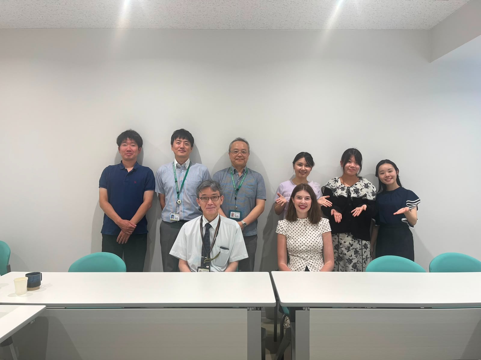
Upon arrival in Japan, specifically at the students' guesthouse, my colleague and I were greeted by Mr. Asahina, an administrative assistant at Hyogo Medical University, who guided us through the students' accommodation as well as the University building. He also provided us with additional information regarding the exchange program. On the first day of the exchange program, my colleague and I met with Mr. Asahina, who escorted us to our respective departments. My department of choice was the Department of Anatomy and Cell Biology. I was immediately introduced to the head of the department, Professor Hideshi Yagi, along with the other professors and staff members. I was also invited to participate in the morning meeting where the professors discussed their research projects. At the end of the meeting, I learned that I would conduct a short research project revolving around the Pacinian corpuscle, a specialized, rapidly adapting mechanoreceptor composed of a central sensory axon surrounded by a dense inner core formed by modified Schwann cells and a perineurial-like outer capsule. Its function is to detect deep pressure and high-frequency vibrations. I was assigned to Instructor Hirosato Kanda, who was my main mentor during the exchange program. Furthermore, I was supervised by the head of the department, Professor Hideshi Yagi.
During those three weeks, I gained valuable insights and acquired a wide range of practical laboratory skills. Throughout my first week, I used a microtome, a mechanical cutting tool, in order to slice extremely thin sections of biological tissue - in this case, those of a Pacinian corpuscle. It was my first time using such an impressive instrument and the whole sectioning process required a lot of precision in order to make satisfactory sections, which were later used for slide preparation. Over the following two weeks, I conducted H&E staining and performed immunohistochemistry utilizing primary antibodies such as DAPI, GLUT1 and S100 in order to evaluate the structural compartmentalization of the Pacinian corpuscle. DAPI mapped the nuclear density across the concentric lamellae of the Pacinian corpuscle due to its binding to DNA, S100 was localized to the modified Schwann cells of the inner core and GLUT1 delineated the perineurial-like outer core and capsule. These primary antibodies were diluted in phosphate-buffered saline (PBS) at various ratios to determine the optimal dilution for maximizing signal intensity and minimizing background noise under a confocal microscope. Confocal microscopy imaging served as the final stage of my research project. I concluded the research by presenting the results to the professors and students at the Department of Anatomy and Cell Biology on my last day of exchange.
To further enrich this academic experience, I had the privilege of attending the NEURO2026 conference in Kobe, where I gained invaluable exposure to the latest advancements in international neuroscience research, which deeply reinforced my commitment to the field. During our time in Kobe, Instructor Hirosato Kanda made sure to show me another facility of Hyogo Medical University – the Kobe Campus, which houses the Schools and Graduate Schools of Pharmacy, Nursing and Rehabilitation. At the Nishinomiya Campus, he also showed me the dissection room, where medical students perform dissections on cadavers under the guidance of their professors as a part of their anatomy course. He also guided me through the main facilities of the Hyogo Medical University Hospital, located on the same grounds.
In the end, I would like to express my utmost gratitude for being able to participate in this exchange program and spend a part of my academic journey at Hyogo Medical University. I would like to thank the professors and the staff of the Department of Anatomy and Cell Biology for their kindness and hospitality towards me. I am especially grateful to Instructor Hirosato Kanda, who patiently guided me through every step of my research project, teaching me invaluable medical insights as well as various aspects of the Japanese medical education system and culture. I would also like to express my sincere appreciation to the head of the department, Professor Hideshi Yagi, whose guidance ensured I always felt supported and welcomed. Furthermore, meeting my medical colleagues, Hitomi, Tamaki and Shiho, was an absolute highlight and I truly value the friendships and professional connections we formed.
Ultimately, I deeply appreciated every aspect of this exchange program. It allowed me to immerse myself in the Japanese medical education system, discover fascinating medical insights and experience Japan's incredible culture, traditions and landmarks. This was truly a once-in-a-lifetime experience that has immensely contributed to both my personal and academic growth and I sincerely hope to visit Japan again in the near future.