About the Corporation
Successful accurate visualization of the internal iliac vein to reduce the risk of bleeding during surgery for local recurrence of rectal cancer - Multidisciplinary research by radiologists and surgeons -
Dr. Wataru Shiromoto, a radiologist in the Department of Radiological Technology Hyogo Medical University Hospital (located in Nishinomiya, Hyogo Prefecture; Director: Hiroki Ikeuchi), in collaboration with Dr. Kei Kimura, lecturer in the Department of Lower Gastrointestinal Surgery, et al., have searched for an appropriate MRI sequence to accurately depict the internal iliac vein, which is a risk factor for surgical treatment of local recurrence of rectal cancer, and published a paper on the accuracy of depiction using the "MRI True-FISP" sequence method.
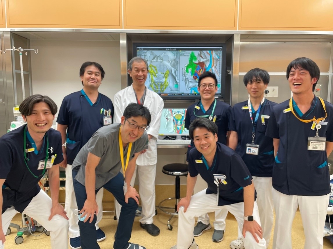
Top row, from left: Takahiro Minamoto (Chief of the Department of Radiological Technology), Masataka Ikeda (Chief Professor of the Department of Lower Gastrointestinal Surgery), Wataru Shiromoto (Department of Radiological Technology), Hotaka Nakagiri (Department of Radiological Technology), Takumu Egashira (Department of Radiological Technology) Bottom row, from left: Masato Kiriki (Department of Radiological Technology), Kei Kimura (Lecturer of the Department of Lower Gastrointestinal Surgery), Daisuke Nakajima (Department of Radiological Technology)
Research Summary
In cases of local recurrence of rectal cancer, damage to the internal iliac vein during treatment can cause fatal bleeding. Therefore, accurate identification of the internal iliac vein before surgery is necessary. In this study, we searched for an appropriate MRI sequence that would accurately depict the internal iliac vein in patients scheduled for surgery for local recurrence of rectal cancer, and succeeded in accurately depicting the internal iliac vein using the "MRI True-FISP" sequence method.
Research Background
In the case of local recurrence of rectal cancer, the layer that was dissected in the initial surgery must be dissected again, making it a highly challenging procedure that makes Laboratory of Anatomy identification difficult. In addition, the branching morphology of the internal iliac vein is extremely complex. Although there have been studies to date that have used CT and MRI to evaluate the first and second branches of the internal iliac vein, there have been no studies that have visualized the peripheral branches, which are the most important in surgery for local recurrence of rectal cancer. In this study, we decided to visualize the internal iliac vein using a sequence called "MRI True-FISP," which is excellent for visualizing cardiovascular vessels.
Research Methods and Results
Research Methodology
Between March 2023 and November 2023, 11 patients who required surgery for local recurrence of rectal cancer were included.
CT was performed using CT angiography with iodine contrast agent, and MRI was performed using the True-FISP method. In addition, gadolinium contrast agent with T1 shortening effect was administered to clearly visualize blood vessels, and anticholinergic drug butylscopolamine was administered to suppress artifacts caused by intestinal peristalsis.
In order to reconstruct blood vessels and pelvic anatomy in 3D for reference during surgery, the internal iliac vessels obtained by CT and the internal iliac vessels obtained by MRI were analyzed using the image analysis system "SYNAPSE VINCENT®︎," and vascular visualization was evaluated by measuring the contrast ratio between the internal iliac vessels and the piriformis muscle, which is the muscle closest to the internal iliac vessels.
Results
The contrast ratio of the internal iliac vein was 0.23 for CT and 0.55 for MRI, with the contrast ratio of the internal iliac vein on MRI being significantly higher (p<0.01).Distortion was evaluated using CT and MRI fusion, and the linear distance from the left and right ischial spines, and from the ischial spine to the coccyx and from the ischial spine to the branching point of the superior gluteal artery was measured. The average error was less than 1mm, and no distortion was observed in the CT and MRI fusion. By superimposing the pelvis and arteries, the images were useful for surgery.
The results of this study suggest that the MRI True-FISP sequence method can accurately depict the peripheral branches of the internal iliac vein.
Future challenges
Because the number of cases in this study was small, it is necessary to collect more cases and compare them with previous cases to consider ways to reduce bleeding.
Source of research funds etc.
none
Paper information
Publication
Magn Reson Imaging. 2024 Apr 7;111:9-14. doi: 10.1016/j.mri.2024.04.006.
Paper title
Delineation of the internal iliac vein using MRI with true FISP sequence in patients with locally recurrent rectal cancer: A pilot study using CT/ MRI fusion
author
Wataru Shiromoto 1, Kei Kimura 2, Masato Kiriki 3, Masashi Koizumi 3, Hotaka Nakagiri 3, Daisuke Nakajima 3, Yusuke Kawanaka 4, Kazuhiro Kitajima 4,
Haruyuki Takagi 4, Naohito Beppu 5, Kozo Kataoka 5, Masataka Ikeda 5, Koichiro Yamakado 4
Affiliation
1 Department of Radiological Technology Hyogo Medical University Hospital
2. Department of Gastroenterological Surgery, Hyogo Medical University
3Department of Radiological Technology Hyogo Medical University Hospital
4Department Department of Radiology Hyogo Medical University College of Medicine
Hyogo Medical University of Medicine, Department of Gastroenterological Surgery, Lower Gastroenterological Surgery