Research Seeds Collection
AI-based image diagnosis support system for facial bone fractures
Information updated: June 12, 2024
- Seeds Information
- Researcher Information
- What do you expect from collaboration with companies?
- Contact for this research
Seeds Information
keyword
CT image database diagnostic support system
Field
Plastic Surgery Database AI
Overview
X-ray CT scans are essential for diagnosing facial bone fractures, but for doctors who are not familiar with them, interpretation of the images is not easy. Facial bone fractures progress to healing within 2-3 weeks after injury, so if a fracture is identified after that time, reduction of the fracture becomes extremely difficult. Therefore, it is important to consult a specialist at a relatively early stage about the suitability of surgery.
Aiming to construct an image diagnosis support system for facial bone fractures, we are collecting CT images of the head and face taken for diagnostic purposes and developing an artificial intelligence system for diagnosing facial bone fractures.
Most facial bone fractures occur on one side, with the other side remaining normal. By accumulating data from the healthy side, we will construct 3D data on the condition of the craniofacial bones of people currently living in Japan.
What's new?
Currently, 3D data on the shape of the skull and facial bones of Japanese people only exists for specific individuals or on a small scale. There are various differences between the craniofacial skeletons of Caucasians and Mongoloids, and for medical treatment in Japan, a system trained on data obtained in Japan is necessary.
What are its advantages over other studies?
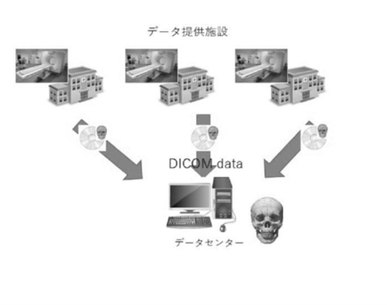
Through this prospective study, we obtained consent, which also included the construction of a 3D database of the craniofacial bones of people living in Japan, and are collaborating with 10 institutions to collect image data taken for the diagnosis of facial bone fractures.
UMIN Study ID: UMIN000039624 "Application of artificial intelligence to image diagnosis of facial bone fractures"
What problem does it help solve?
If an AI system could be used to assist in the diagnosis of facial bone fractures, it could improve the accuracy of diagnosis and reduce the number of oversights, even for non-specialists. It would also enable automatic diagnostic assistance from remote locations, which is expected to improve the quality of life of injured people and promote their rehabilitation into society.
Possibility of other applications and developments
Facial bone fractures are often unilateral, with the other side remaining healthy. By collecting healthy parts, we can explore what kind of facial skeleton modern Japanese people have.
By accumulating data on an unprecedented scale, we can construct a standard skeletal model of modern Japanese people, which will serve as a basis for examining whether the skeletons of future Japanese people will change due to the influence of diet and the environment. It will also enable cultural anthropological studies, such as comparisons with people buried in the past and with other races.
It will also enable computer simulations such as finite element method models to be performed using data from Japanese people, and will also be useful industrially, for example in the design of glasses, various wearable devices, helmets, hats, and other items.
Related Patents
―
Related papers
- Automatic landmark prediction in craniofacial CT images: Three dimensional coordinates prediction of landmarks on craniofacial bone. Nishimoto S.JSAI2021(0)4I4GS7e04, 2021
- Three-dimensional cranio-facial landmark detection in CT slices from a publicly available database, using multi-phased regression networks on a personal computer. Nishimoto S, Ishise H, et al. medRxiv2021.03.21.21253999,2021
- Machine learning-based noise reduction for craniofacial bone segmentation in CT images. Nishimoto S, Ishise H, et al. medRxiv2022.06.26.22276925,2022
- Three-Dimensional Craniofacial Landmark Detection in Series of CT Slices Using Multi-Phased Regression Networks. Nishimoto S, Ishise H, et al. Diagnostics, 13, 1930, 2023
Reference Chart
Researcher Information
| full name | Satoshi Nishimoto |
|---|---|
| Affiliation | School of Medicine Department of Plastic Surgery |
| Specialization | Plastic Surgery Craniofacial Surgery |
| Collaborative Researcher | Hisako Ishise, Toshihiro Fujiwara, Kenichiro Kawai, Masao Kakibuchi, and Takaaki Nakajima |
| Related links | Course introduction website |
What do you expect from collaboration with companies?
・Cooperation in constructing a standard skeletal model of modern Japanese people
・Improvement of the accuracy of image diagnostic support systems for facial bone fractures
Contact for this research
兵庫医科大学 大学事務部 研究推進課
E-mail: chizai@hyo-med.ac.jp
Tel: 0798-45-6488